
The goal of the Vásquez lab is to understand the molecular rules that cells use to build and shape functional organs. Currently, we are studying the morphogenesis of the Drosophila melanogaster renal system – the Malpighian tubules. The ability to watch this organ grow and manipulate it in real time make it an ideal system for dissecting the emergent properties cells use to build complex tissue structures.
research
The Vásquez lab seeks to identify the molecular and physical mechanisms that cells use to build our organs. The goal of our lab is to dissect the emergent properties cells used to build complex higher-order tissue structures, like our organs. We take a multidisciplinary approach, using a combination of quantitative light microscopy, genetics, and biochemistry using the developing renal system of the fruit fly (Drosophila melanogaster Malpighian tubules) as an in vivo model for organogenesis.
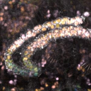
The developing fly renal system involves the generation and extension of two pairs of tubes that fold on themselves in a stereotypic manner providing an ideal model in which to isolate the parameters that generate specific cell shapes while keeping the cells in an in vivo context. Work in our lab will leverage the known pathways and molecular mechanisms at work in studying cells at an individual basis to uncover how cells integrate these features into highly regulatable 3D tissue forms.

Current work in our lab will focus around two central questions:
[1] How do cells manage changing shape?
Over the course of 6 hours Malpighian tubules extend 4-fold in length without any cell proliferation. Throughout this process, tubule cells transition from tall columnar cells to short cuboidal cells. This transformation provides an opportunity to understand how cellular domains adapt to changing cellular dimensions in a developing organ.

[2] How do cells build tubes that fold on themselves and still function?
We are beginning to understand how tissues make prevalent epithelial motifs like folds and tubes. However, the tissue structures that make up organs are more complicated than these simple motifs. These motifs are combined to generate higher order structures with 3D folded patterns, for example a tube that loops, or a fold within the epithelial structure of a tube.

news
Hayden passes her general exam, pumpkins galore, and more!
Julene joins the lab Julene is a third year undergraduate student, she’ll be working with Michelle to test out perturbations to tubule cell number. We’re excited to have her on board. Michelle attends & presents at the AI & Digital Biology Symposium Michelle attended and presented at this local conference hosted by our neighbors at …
Continue reading “Hayden passes her general exam, pumpkins galore, and more!”
summer time the living’s easy + TWO rotation students!
2nd annual Golden Gardens picnic with the Kwon Lab We had a lovely afternoon picnic with the Kwon lab at Golden Gardens. TWO rotation students The lab welcomes two rotation students this fall quarter: Gavin Wheeler & Val Browning! We are super excited to have them both in the lab with us! Marcel updates and …
Continue reading “summer time the living’s easy + TWO rotation students!”
bowling, the alps, graduates, and more! 🎳 🏔️🎓
Megan presents a poster at the Undergraduate Research Symposium Megan had the opportunity to present her work to the broader UW community. After presenting her work at NWSDB earlier this year, this presentation was easy as pie! 1st inaugural “hang” with the Akamatsu Lab 🎳 After some intense cat herding by Valerie and Vesta – …
Continue reading “bowling, the alps, graduates, and more! 🎳 🏔️🎓”
people
chronological order

Claudia Vásquez

Michelle Chicas

Vesta Baumgartner

Megan Yi

Hayden Gizinski

Isabel Mejia Natividad

Julene Wang

Israfel Ibrahim

Kazuma Fukuda

Marcel the Shell with Shoes On
ALUMNI
reverse chronological order
| name | role in Vásquez lab | next stop |
| Gavin Wheeler | rotation student Fall 2025 | next rotation! |
| Val Browning | rotation student Fall 2025 | next rotation! |
| Rishik Chakrabarti | high school intern 2023-2025 | Michigan State University |
| Jade Gibbs | undergraduate researcher Winter & Spring 2025 | graduation! |
| Gabe Landau | undergraduate researcher Winter 2025 | graduation! |
| Makenna Carnahan | undergraduate researcher Winter 2024 – Fall 2024 | Suzie Pun’s lab in Bioengineering |
| Melissa Calderón Bracho | BRIGHT UP summer high school student 2023 and 2024 | Claremont McKenna College |
| Lily Worst | rotation student Winter 2024 | graduate student in Chip Asbury lab |