The Vásquez lab seeks to identify the molecular and physical mechanisms that cells use to build our organs. The goal of our lab is to dissect the emergent properties cells used to build complex higher-order tissue structures, like our organs. We take a multidisciplinary approach, using a combination of quantitative light microscopy, genetics, and biochemistry using the developing renal system of the fruit fly (Drosophila melanogaster Malpighian tubules) as an in vivo model for organogenesis.
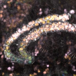
The developing fly renal system involves the generation and extension of two pairs of tubes that fold on themselves in a stereotypic manner providing an ideal model in which to isolate the parameters that generate specific cell shapes while keeping the cells in an in vivo context. Work in our lab will leverage the known pathways and molecular mechanisms at work in studying cells at an individual basis to uncover how cells integrate these features into highly regulatable 3D tissue forms.

Current work in our lab will focus around two central questions:
[1] How do cells manage changing shape?
Over the course of 6 hours Malpighian tubules extend 4-fold in length without any cell proliferation. Throughout this process, tubule cells transition from tall columnar cells to short cuboidal cells. This transformation provides an opportunity to understand how cellular domains adapt to changing cellular dimensions in a developing organ.

[2] How do cells build tubes that fold on themselves and still function?
We are beginning to understand how tissues make prevalent epithelial motifs like folds and tubes. However, the tissue structures that make up organs are more complicated than these simple motifs. These motifs are combined to generate higher order structures with 3D folded patterns, for example a tube that loops, or a fold within the epithelial structure of a tube.