 |
Claudia Vasquez
Assistant Professor of Biochemistry BA, BS 2010, Brandeis University
PhD 2015, Massachusetts Institute of Technology
|
Honors
- 2023 Presidential Membership Initiative, Genetics Society of America
- 2021 Intersections Science Fellow
- 2021 Stanford.Berkeley.UCSF Next Generation Faculty Symposium Fellow
- 2021 ASAPBio Fellow
- 2020 Leading Edge Fellow
- 2010 Phi Beta Kappa Scholar
- 2009-2010 Schiff’s Fellow, Brandeis University
- 2006-2010 Justice Louis D Brandeis Scholar, Brandeis University
Research
What are the molecular and physical mechanisms that cells use to build our organs? The goal of our lab is to dissect the emergent properties cells used to build complex higher-order tissue structures, like our organs. We take a multidisciplinary approach, using a combination of quantitative light microscopy, genetics, and biochemistry using the developing renal system of the fruit fly (Drosophila melanogaster Malpighian tubules) as an in vivo model for organogenesis.
The developing fly renal system involves the generation and extension of two pairs of tubes that fold on themselves in a stereotypic manner providing an ideal model in which to isolate the parameters that generate specific cell shapes while keeping the cells in an in vivo context. Work in our lab will leverage the known pathways and molecular mechanisms at work in studying cells at an individual basis to uncover how cells integrate these features into highly regulatable 3D tissue forms.
Current work in our lab will focus around two central questions:
-
- How do cells manage changing shape?Over the course of 6 hours Malpighian tubules extend 4-fold in length without any cell proliferation. Throughout this process, tubule cells transition from tall columnar cells to short cuboidal cells. This transformation provides an opportunity to understand how cellular domains adapt to changing cellular dimensions in a developing organ.
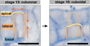
Tubule cells change shape during organogenesis. Confocal images of tubule cells at representative stages immunostained for a junctional protein (Ssk). Scale bars, 5 µm
- Put simply, how do cells build tubes that fold onto themselves and still function?Over the course of 6 hours Malpighian tubules extend 4-fold in length without any cell proliferation. Throughout this process, tubule cells transition from tall columnar cells to short cuboidal cells. This transformation provides an opportunity to understand how cellular domains adapt to changing cellular dimensions in a developing organ.
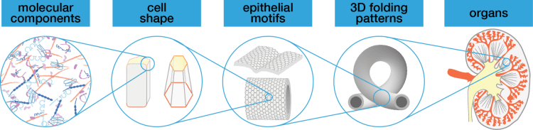
From molecules to organs – how do cells organize themselves and with each other to build complex and functional structures?
We can begin to dissect how cells build different complex tissue patterns by comparing the strategies cells used to build the anterior and posterior tubules – which have dramatically different morphologies. What is different about the anterior and posterior tubule cells and their environments – how do these differences contribute to organ shape and function?
- How do cells manage changing shape?Over the course of 6 hours Malpighian tubules extend 4-fold in length without any cell proliferation. Throughout this process, tubule cells transition from tall columnar cells to short cuboidal cells. This transformation provides an opportunity to understand how cellular domains adapt to changing cellular dimensions in a developing organ.
We expect that there will be a diversity of strategies that cells and tissues use to generate 3D tissue structures. To this end, our work will initially focus on the Drosophila renal system and use that as a launching pad to study other organs in Drosophila and other organisms, with the goal of building towards a molecular scale understanding of how cellular shape feeds into organ form and function.
Publications: