The best cell biologists on earth are not people — they are microbes!
• Neisseria gonorrhoeae and Neisseria meningitidis
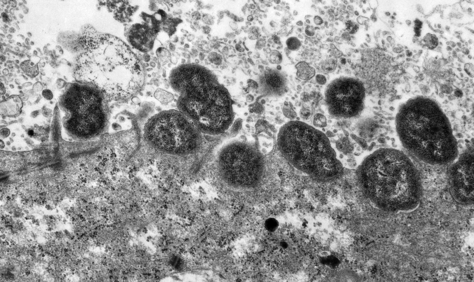
After a long hiatus we’ve re-started our work on bacteria that cause gonorrhea and meningitis. As a grad student with Maggie So (then at OHSU), Alex worked on the type 4 pilus, and on how host cells respond when Neisseria adhere. Type 4 pili are among the most remarkable machines in nature. Acting as grappling hooks, they can feed out a filament, attach to a surface, and reel the filament in, like a grappling hook. In collaboration with Mike Sheetz, we were perhaps the first to directly observe pilus retraction. At about the same time, Jeff Skerker and Howard Berg used total internal reflection microscopy to watch the type 4 pili of Pseudomonas extending and retracting.
- Merz AJ, So M and Sheetz MP. 2000. Pilus retraction powers bacterial twitching motility. Nature 407:98-102. –pdf
- Merz AJ and Forest KT. 2002. Review: Bacterial surface motility: slime trails, grappling hooks, and nozzles. Current Biology 12:R297-303. –pdf
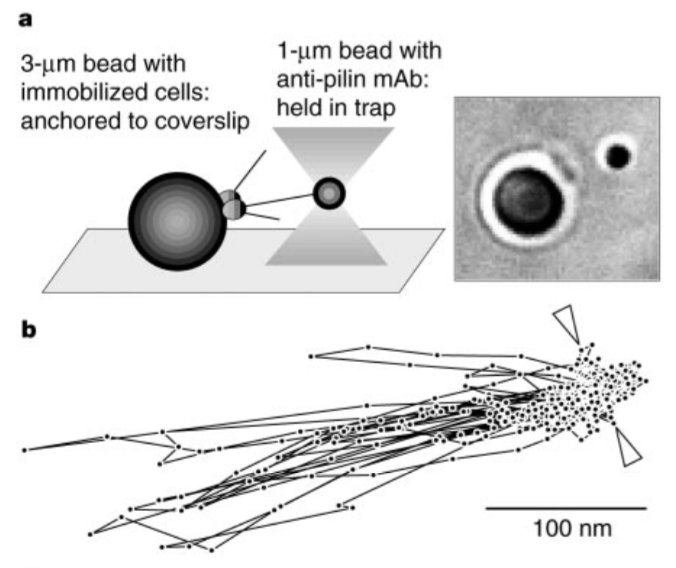
In the experiment shown below, laser tweezers are used to hold a small bead near bacteria which are anchored to a larger bead. The bacterial pili (not visible by transmitted light microscopy) reach out and repeatedly pull the bead out of the optical trap, toward the bacteria.
 |
Here’s an animation depicting how the type 4 pilus might extend and retract, from the Jensen Lab:
In other work, we found that the type 4 pili of Neisseria species allow the bacteria not only to adhere to host cells, but trigger a remarkable series of cytoskeletal and membrane rearrangements in those cells. These rearrangements depend on pilus retraction, indicating that mechanical force is part of the signalling process. Pili also increase the ability of the bacteria to invade into deeper tissues.

- Merz AJ, Enns CA and So M. 1999. Type IV pili of pathogenic Neisseriae elicit cortical plaque formation in epithelial cells. Molecular Microbiology 32:1316-1332. doi: 10.1046/j.1365-2958.1999.01459.x
- Merz AJ and So M. 2000. Review: Interactions of pathogenic Neisseriae with epithelial cell membranes. Annual Review of Cell & Developmental Biology 16:423-457. –pdf
Now we are working to understand the biochemical mechanisms used by these pathogens to assemble and retract pili, and what happens when they attach to host cells. In our current experiments we’re focusing on biochemical approaches.
• Bacterial secreted effectors
Alex got interested in bacterial secreted effectors in grad school. These are bacterial proteins injected from bacteria into other cells. Over the last decade we’ve collaborated with the Mougous group here at UW on studies of bacterial type VI effectors used to kill other bacteria, and to manipulate host cells. Currently, we are using bacterial effectors as tools in our studies of membrane trafficking.
- Hood RD, Singh P, Hsu F, Guevener T, Carl MA, Trinidad RRS, Silverman JM, Ohlson BB, Hicks KG, Plemel RL, Li M, Schwartz S, Wang WY, Merz AJ, Goodlett D, Mougous J. 2010. Discovery of Type VI secretion substrates in Psudomonas aeruginosa: identification of a toxin targeted to bacteria. Cell Host & Microbe, doi: 10.1016/j.chom.2009.12.007 *F1000 Must Read Paper.
- Ledvina HD, Cundiff JA, Kelly, KA, Plemel RL, Eshraghi A, Brook S, Peterson SB, Lee B, Alder M, Merz AJ, Skerrett SJ, Celli J, Mougous JD. 2018. A phosphatidylinositol 3-kinase effector alters phagosomal trafficking to promote intracellular growth of Francisella. Cell Host & Microbe, doi: 10.1016/j.chom.2018.07.003. A Preview featuring this paper has been published: doi: 10.1016/j.chom.2018.07.016
 |
 |
• Virology
We have thoughts. Perhaps in the near future, results. Time will tell!